For scientists all over the place, April 25 is promising. It is DNA Day and remembers the date in 1953 when researchers Francis Crick, Rosalind Franklin, James Watson and Maurice Wilkins distributed original logical papers portraying the helical design of the DNA particle. In 2003, April 25 was utilized to report the culmination of the Human Genome Project. Presently yearly merriments on this day commend the atom of existence with new revelations. What better opportunity to give another image of DNA.
I’m DNA DAVE (or if nothing else my tag beginning around 1984 says as much), and something my lab likes to do is to “see” DNA. We take pictures of DNA so we can straightforwardly gauge things that are hard to measure utilizing aberrant strategies that normally include sequencing the four synthetic units of DNA, called bases.
For instance, I might want to know where on every chromosome the course of DNA replication starts. Blunder free duplication of DNA is fundamental for delivering solid cells. At the point when this cycle is inadequate or upset, the outcome can cause malignant growth and different sicknesses.
In our picture that recognizable twofold helix flight of stairs isn’t noticeable on the grounds that this viewpoint is zoomed out – like taking a gander at the guide of a country versus a city. Likewise every one of these particles is identical to 50,000 turns of the helical flight of stairs – a significant fragment of a human chromosome.
Making a guide of DNA
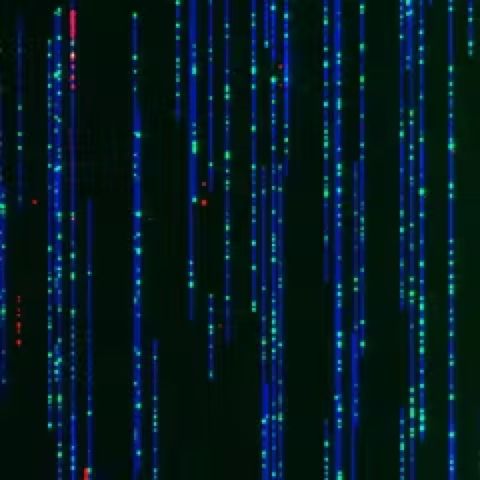
This picture, taken with a gadget called the Bionano Genomics Saphyr imager, highlights individual DNA particles – shaded in blue, green and red. These strands of DNA have been adjusted by stringing them through restricted tubes – called nanochannels – that fit just a single piece of DNA. As the DNA slips into the cylinder, the strands fix.
The entire DNA atom is hued blue and the green tick marks are tourist spots – or explicit arrangements of DNA that happen on normal each 4,500 base matches. The example of milestones give a special finger impression which lets us know where we are along the length of a chromosome. The red fluorescent blips label the places where the DNA has started to recreate. These locales are classified “beginnings of replication” and are where the DNA initially loosens up so the duplication cycle can begin.
Scientists at Bionano Genomics in San Diego fostered this nanochannel innovation to graph areas of chromosomes that were generally unmappable, because of precarious hereditary groupings that make it challenging to decide the request for the four bases. This gadget tackled the issue by “looking” at the plan of successions on each particle in turn and can peruse 30 billion base sets in a single hour – what could be compared to 10 human genomes.
My group and that of Nick Rhind at the University of Massachusetts perceived that this nanochannel innovation would permit us to lead an examination never endeavored: map every one of the places where DNA replication starts at the same time on large number of single DNA strands.

Before a cell can partition into two free cells, the DNA should make a duplicate of itself so every one gets a total arrangement of chromosomes. To comprehend how the hereditary material is copied it is crucial for know where along the chromosome the cycle starts. That has been the best test to concentrating on how the replication of our own chromosomes happens and subsequently the thing is turning out badly in such countless sicknesses, similar to malignant growth, in which replication turns out badly.
DNA replication and malignant growth
Beginnings of replication have been slippery on the grounds that they happen at many destinations on various particles so we really want to take a gander at single DNA atoms to recognize them. Despite the fact that researchers have had the option to see single DNA particles since the mid 1960s, we were unable to tell where in the chromosomes any atom came from so we were unable to plan anything.
Kyle Klein, a Ph.D. understudy in my lab, named residing human immature microorganisms with red fluorescent atoms that undeniable places where DNA replication was occurring, which were planned with the Bionano gadget. These pictures were then superimposed onto the blue and green DNA guides of similar DNA particles.
We anticipate that this strategy should totally change how we might interpret how human chromosomes imitate. Besides, since most chemotherapy drugs for malignant growth therapy and most cancer-causing agents – or disease causing synthetic compounds – in our current circumstance work by going after DNA when it duplicates, we anticipate that this strategy should give a quick and far reaching test for how these synthetic compounds disturb DNA replication. We additionally trust it uncovers how we could lighten these unfortunate results, and how we could foster better and less harmful chemotherapy medicines.